Structural and functional study of ACAP1 - a key component of a clathrin complex for endocytic recycling
(In collaboration with Prof. Victor Hsu, Harvard Medical School)
1. Structural elucidation of cargo-binding auto-inhibition mechanism of ACAP1

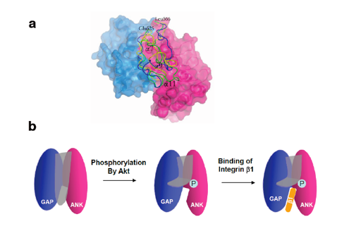
Structural elucidation of cargo-binding auto-inhibition mechanism of ACAP1. (a) Crystal structure of GAP-ANK domain of ACAP1. GAP domain is colored by blue and ANK domain by pink. The linker region between GAP and ANK domain is modeled with green for WT, yellow for S554D and blue for S554A. (b) A working model for the auto-inhibition mechanism of ACAP1. which can be regulated by phosphorylation of the linker region.
Coat complexes mediate the proper sorting of protein cargoes among intracellular membrane compartments. An emerging class of coat components has been the GTPase activating proteins (GAPs) that act on the ADP-Ribosylation factor (ARF) family of small GTPases. ACAP1 is an ARF6 GAP that also acts as a key component of a clathrin complex for endocytic recycling. Phosphorylation of ACAP1 by Akt has been shown to enhance cargo binding of integrin beta1 for stimulation-dependent integrin recycling. Our structural studies found that this phosphorylation relieves a local mechanism of inhibition, which involves a linker region acting to obstruct the cargo-binding site in ACAP1. Moreover, cargo binding involves the GAP domain, which reveals an unexpected role for this catalytic domain as an ARF effector. These findings advance a mechanistic understanding of how an ARF GAP acts in cargo binding, and also shed molecular insight into a key regulatory mechanism that controls integrin recycling.
Reference:
Bai M., Pang X., Lou J., Zhou Q., Zhang K., Ma J., Li J., Sun F.*, Hsu V.* (2012), Mechanistic insights into regulated cargo binding by ACAP1. Journal of Biological Chemistry,287: 28675-28685.
2. A novel membrane remodeling mechanism by the BAR-PH domain of ACAP1
(In collaboration with Prof. Victor Hsu, Harvard Medical School; Prof. Jun Fan, City University of HongKong)

Molecular detail of how ACAP1 achieves membrane remodeling.Top-left, negative-stain electron microscopy visualizing liposomes incubated with BAR-PH domain of ACAP1. Top-right, cryoEM map of a liposomal tubule coated with BAR-PH domains of ACAP1. Bottom-right, structural model of BAR-PH helical assembly on liposomal tubule. Bottom-left, the final simulated model of PH domain engaging into the membrane.
The BAR (Bin-Amphiphysin-Rvs) domain undergoes dimerization to produce a curved protein structure, which superimposes onto membrane through electrostatic interactions to sense and impart membrane curvature. In some cases, a BAR domain also possesses an amphipathic helix that inserts into the membrane to induce curvature. ACAP1 (Arfgap with Coil coil, Ankyrin repeat, and PH domain protein 1) contains a BAR domain. Here, we show that this BAR domain can neither bind membrane nor impart curvature, but instead requires a neighboring PH (Pleckstrin Homology) domain to achieve these functions. Specific residues within the PH domain are responsible for both membrane binding and curvature generation. The BAR domain adjacent to the PH domain instead interacts with the BAR domains of neighboring ACAP1 proteins to enable clustering at the membrane. Thus, we have uncovered the molecular basis for an unexpected and unconventional collaboration between PH and BAR domains in membrane bending.
Reference:
Pang X., Fan J., Zhang Y., Zhang K., Gao B., Ma J., Li J., Deng Y., Zhou Q., Egelman E.H., Hsu V.W.* and Sun F.* (2014), A PH Domain in ACAP1 Possesses Key Features of the BAR Domain in Promoting Membrane Curvature. Developmental Cell, 31(1) : 73-86. doi: 10.1016/j.devcel.2014.08.020.
Chan C. Pang X., Zhang Y., Niu T., Yang S., Zhao D., Li J., Lu L., Hsu, V.W., Zhou J.*,Sun F.* and Fan J.* (2019), ACAP1 assembles into an unusual protein lattice for membrane deformation through multiple stages. PLOS Computational Biology,15 (7): e1007081. doi: 10.1371/journal.pcbi.1007081.
附件下载: