Structural study of rabbit hemorrhagic disease virus
1. Cryo-electron microscopy reconstructions of two types of wild rabbit hemorrhagic disease viruses.(In collaborations with Prof. Dong Zheng, Beijing Normal University)
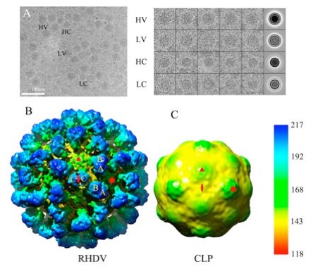
Rabbit hemorrhagic disease was described in China in 1984 and can cause hemorrhagic necrosis of the liver within two or three days after infection. The etiological agent, rabbit hemorrhagic disease virus (RHDV), belongs to the Lagovirus genus in the Caliciviridae family. Compared to other calicivirus, such as rNV and SMSV, the structure of Lagovirus members is not well characterized. In our research, structures of two types of wild RHDV particles, the intact virion and the core-like particle (CLP), were reconstructed by cryo-electron microscopy at 11 ? and 17 ?, respectively. This is the first time the 3D structure of wild caliciviruses CLP has been provided, and the 3D structure of intact RHDV virion is the highest resolution structure in Lagovirus. Comparison of the intact virion and CLP structures clearly indicated that CLP was produced from the intact virion with the protrusion dissociated. In contrast with the crystal structures of recombinant Norovirus and San Miguel sea lion virus, the capsomers of RHDV virion exhibited unique structural features and assembly modes. Both P1 and P2 subdomains have interactions inside the AB capsomer, while only P2 subdomains have interaction inside CC capsomer. The pseudo atomic models of RHDV capsomers were constructed by homology modeling and density map fitting, and the rotation of RHDV VP60 P domain with respect to its S domain, compared with SMSV, was observed. Collectively, our cryo-electron microscopic studies of RHDV provide close insight into the structure of Lagovirus, which is important for functional analysis and better vaccine development in the future.
Reference:
Hu Z., Tian X., Zhai Y., Xu W., Zheng D., Sun F.* (2010), Cryo-Electron Microscopy Reconstructions of Two Types of Wild Rabbit Hemorrhagic Disease Viruses Characterized the Structural Features of Lagovirus. Protein & Cell, 1(1):48-58.
2. Atomic model of rabbit hemorrhagic disease virus by cryo-electron microscopy and Crystallography. (In collaboration with Prof. Hai Pang from Tsinghua University, Prof. Klaus Schulten from University of Illinois at Urbana-Champaign, Prof. Timothy S. Baker from University of California-San Diego, Prof. Dong Zheng from Beijing Normal University and Dr. Jiasen Liu from Harbin Veterinary Research Institute.)
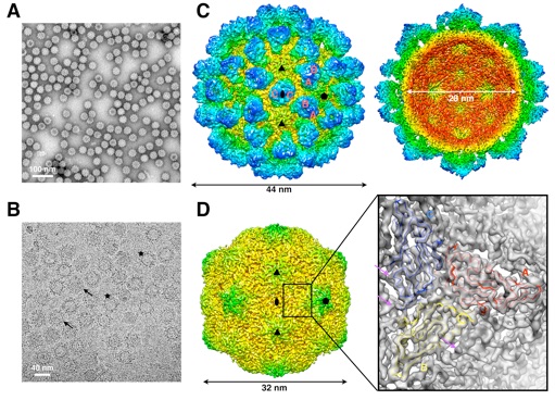
Rabbit hemorrhagic disease, first described in China in 1984. causes hemorrhagic necrosis of the liver. Its etiological agent, rabbit hemorrhagic disease virus (RHDV), belongs to the Lagovirus genus in the family Caliciviridae. The detailed molecular structure of any lagovirus capsid has yet to be determined. Here, we report a cryo-electron microscopic (cryoEM) reconstruction of wild-type RHDV at 6.5 ? resolution and the crystal structures of the shell (S) and protruding (P) domains of its major capsid protein, VP60. each at 2.0 ? resolution. From these data we built a complete atomic model of the RHDV capsid. VP60 has a conserved S domain and a specific P2 sub-domain that differs from those found in other caliciviruses. As seen in the shell portion of the RHDV cryoEM map, which was resolved to ~5.5 ?, the N-terminal arm domain of VP60 folds back onto its cognate S domain. Sequence alignments of VP60 from six groups of RHDV isolates revealed seven regions of high variation that could be mapped onto the surface of the P2 sub-domain and suggested three putative pockets might be responsible for binding to histo-blood group antigens. A flexible loop in one of these regions was shown to interact with rabbit tissue cells and contains an important epitope for anti-RHDV antibody production. Our study provides a reliable, pseudo-atomic model of a Lagovirus and suggests a new candidate for an efficient vaccine that can be used to protect rabbits from RHDV infection.
Reference:
Wang X., Xu F., Liu J., Gao B., Liu Y., Zhai Y., Ma J., Zhang K., Baker T.S., Schulten K., Zheng D.*, Pang H.* and Sun F.*. (2013) Atomic model of rabbit hemorrhagic disease virus by cryo-electron microscopy and crystallography. PLoS Pathogens 9(1): e1003132. doi: 10.1371/journal.ppat.1003132.
附件下载: