Determining the target protein localization in 3D using the combination of FIB-SEM and APEX2
(In collaboration with Center for Biological Imaging, Institute of Biophysics, CAS)
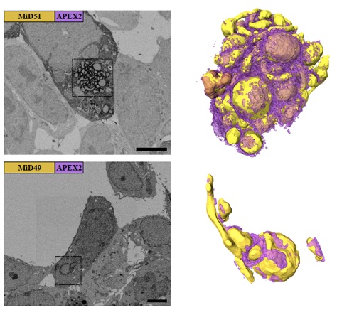
Visualizing the staining pattern in 3D by FIB-SEM. The left columns display the representative SEM micrographs from each volume EM data, and the right columns display the 3D rendering of boxed areas of the left with the yellow spheres for mitochondria and purple layers for APEX2 induced EM contrast.
Determining the cellular localization of proteins of interest at nanometer resolution is necessary for elucidating their functions. Besides super-resolution fluorescence microscopy, conventional electron microscopy (EM) combined with immunolabeling or clonable EM tags provides a unique approach to correlate protein localization information and cellular ultrastructural information. However, there are still rare cases of such correlation in three dimensional (3D) spaces. Here, we developed an approach by combining the focus ion beam scanning electron microscopy (FIB-SEM) and a promising clonable EM tag APEX2 (an enhanced ascorbate peroxidase 2) to determine the target protein localization within 3D cellular ultrastructural context. We further utilized this approach to study the 3D localization of mitochondrial dynamics related proteins (MiD49/51, Mff, Fis1 and Mfn2) in the cells where the target proteins were overexpressed. We found that all the target proteins were located at the surface of the mitochondrial outer membrane accompanied with mitochondrial clusters. Mid49/51, Mff and hFis1 spread widely around the mitochondrial surface while Mfn2 only existed at the contact sites.
Reference:
Shi Y., Wang L., Zhang J., Zhai Y., and Sun F.* (2017), Determining the target protein localization in 3D using the combination of FIB-SEM and APEX2. Biophysics Reports, (in press). doi: 10.1007/s41048-017-0043-x
The supplementary movies (serial slices of cell) of this work can be downloaded here.
附件下载: