In situ protein micro-crystal fabrication by cryo-FIB for electron diffraction
(In collaboration with Center for Biological Imaging, Institute of Biophysics, CAS)
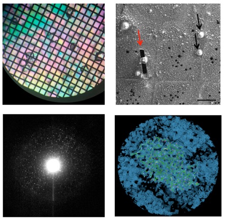
Workflow of in situ cryoFIB-MicroED technique. The protein is crystallized directly on non-magnetic nickel EM grid (top left). Then the grid is vitrified and subjected into cryo focused ion beam fabrication (top right). The thinned cryo-lamella of the protein crystal can diffract electron using 200kV transmission electron microscope (bottom left). Then the crystal structure can be solved from a dataset of electron diffraction of the crystal cryo-lamella.
Micro-electron diffraction (MicroED) is an emerging technique to use cryo-electron microscope to study the crystal structures of macromolecule from its micro-/nano-crystals, which are not suitable for conventional X-ray crystallography. However, this technique has been prevented for its wide application by the limited availability of producing good micro-/nano-crystals and the inappropriate transfer of crystals. Here, we developed a complete workflow to prepare suitable crystals efficiently for MicroED experiment. This workflow includes in situ on-grid crystallization, single-side blotting, cryo-focus ion beam (cryo-FIB) fabrication, and cryo-electron diffraction of crystal cryo-lamella. This workflow enables us to apply MicroED to study many small macromolecular crystals with the size of 2–10 μm, which is too large for MicroED but quite small for conventional X-ray crystallography. We have applied this method to solve 2.5 ? crystal structure of lysozyme from its micro-crystal within the size of 10?×?10?×?10 μm3. Our work will greatly expand the availability space of crystals suitable for MicroED and fill up the gap between MicroED and X-ray crystallography.
Reference:
Li X., Zhang S., Zhang J. and Sun F.* (2018), In situ protein micro-crystal fabrication by cryo-FIB for electron diffraction. Biophysics Reports 4(6): 339-347. doi: 10.1007/s41048-018-0075-x.
The raw data and data processing protocol can be obtained here.
The supplementary movie (continuous electron diffraction pattern ) of this work can be downloaded here.
附件下载: