In situ structural insights into the excitation-contraction coupling mechanism of skeletal muscle
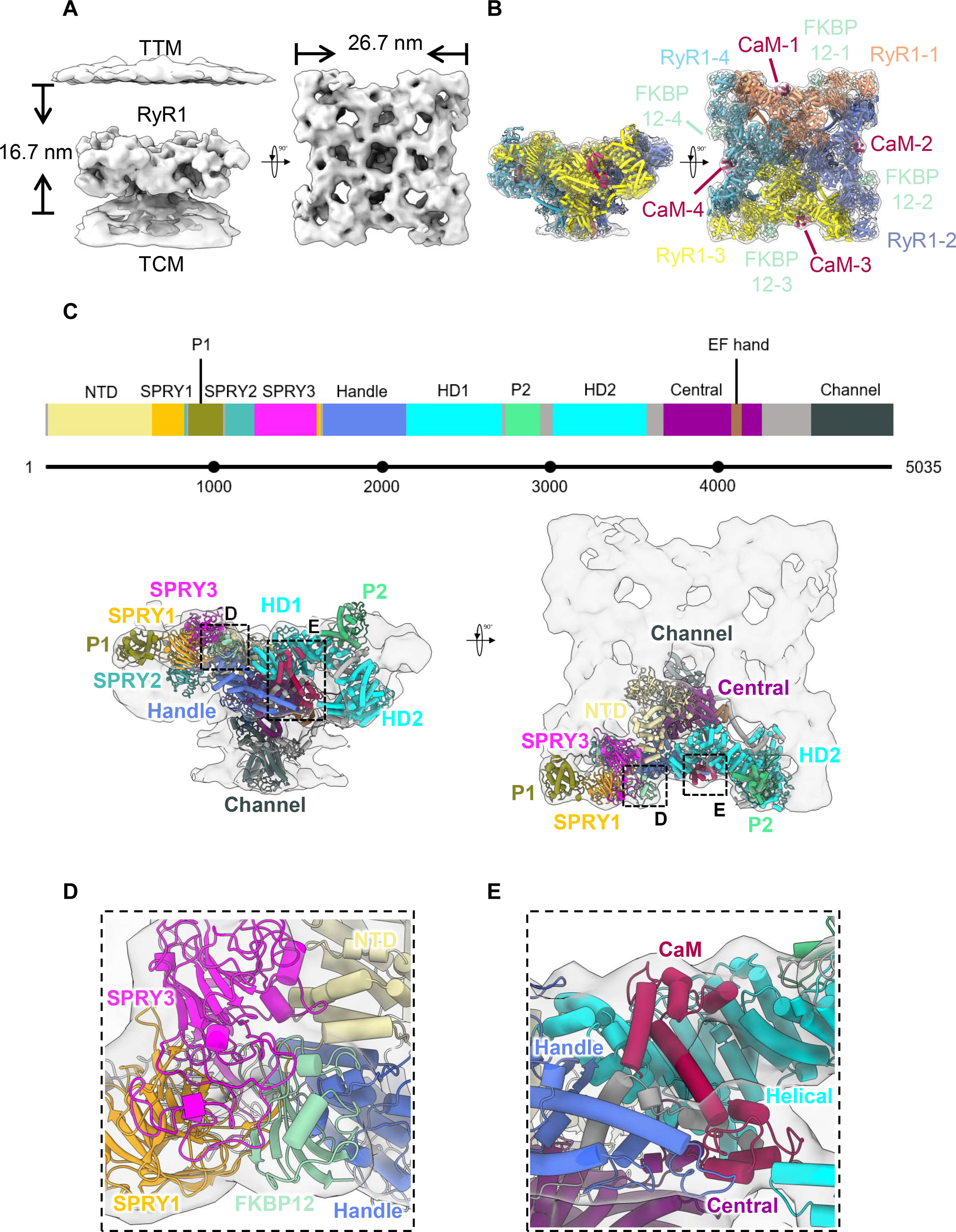
Excitation-contraction coupling (ECC) is a fundamental mechanism in control of skeletal muscle contraction and occurs at triad junctions, where dihydropyridine receptors (DHPRs) on transverse tubules sense excitation signals and then cause calcium release from the sarcoplasmic reticulum via coupling to type 1 ryanodine receptors (RyR1s), inducing the subsequent contraction of muscle filaments. However, the molecular mechanism remains unclear due to the lack of structural details. Here, we explored the architecture of triad junction by cryo–electron tomography, solved the in situ structure of RyR1 in complex with FKBP12 and calmodulin with the resolution of 16.7 Angstrom, and found the intact RyR1-DHPR supercomplex. RyR1s arrange into two rows on the terminal cisternae membrane by forming right-hand corner-to-corner contacts, and tetrads of DHPRs bind to RyR1s in an alternating manner, forming another two rows on the transverse tubule membrane. This unique arrangement is important for synergistic calcium release and provides direct evidence of physical coupling in ECC.

In situ structural insights into the excitation-contraction coupling mechanism of skeletal muscle
附件下载: