1995 - 2000 中国科学技术大学生物系,大学本科
2000 - 2007 中国科学院研究生院认知科学重点实验室,博士研究生
2000 - 2007 中国科学院研究生院认知科学重点实验室,助研
2007 - 2009 中国科学院生物物理研究所脑与认知科学国家重点实验室,博士后
2008 - 至今 中国科学院生物物理研究所脑与认知科学国家重点实验室,副研究员
2012年 “中国科学院卢嘉锡青年人才奖”。
2011年 被研究所推荐成为首批“中国科学院青年创新促进会”会员。
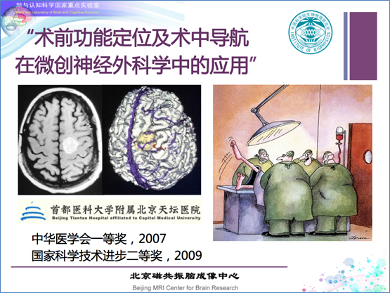
2009年 “国家科技进步奖”二等奖(第四完成人)—“颅脑术中脑认知功能保护的微创神经外科学基础研究与临床应用”。
2007年 “中华医学科技奖”一等奖—“微创神经外科技术平台建立及术中神经功能保护研究”。
认知科学基础理论研究领域,主要研究方向为视知觉大脑左右半球功能不对称性、大范围几何不变性质知觉与视野关系的行为学和神经表达研究(脑成像)。其中大脑半球研究的代表性成果发表在2007年《美国科学院院刊》(作为第一作者和共同通讯作者),并为当期的“本期导读”所介绍。
2002年始作为主要成员参加了以国内首台科研专用的3T磁共振系统、国内唯一一台人类全身7T磁共振系统为核心的、与国际接轨的脑成像研究平台建设。并借助此平台,自2005年开始开展了一系列和临床医院的转化医学合作研究,同天坛医院、同仁医院、宣武医院等著名脑系科医院,合作进行功能成像技术在临床诊断、手术治疗前准备,以及临床基础研究中的应用探索。
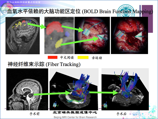
1. 与天坛医院神经外科开展认知科学研究实验范式和功能磁共振技术对手术、患者脑功能保护的转化研究:课题“中国人语言区术前皮层功能定位”课题上的合作,累计完成了500多例次病例采集;课题“颅脑术中脑认知功能保护的微创神经外科学基础研究与临床应用”获2009年“国家科技进步奖”二等奖(个人排名第4);课题“微创神经外科技术平台建立及术中神经功能保护研究”获2007年“中华医学科技奖”一等奖。相关方法应用已推广至“中国胶质瘤协作组”,以及天坛医院放射科、宣武医院功能神经外科、清华大学第二附属医院癫痫中心、中国人民解放军304医院神经外科、海淀医院功能神经科等临床单位,为术中对患者重要脑功能进行有效的保护,降低患者术后致残率和致残级别,提高患者术后康复水平和生活质量,起到了重要的作用。
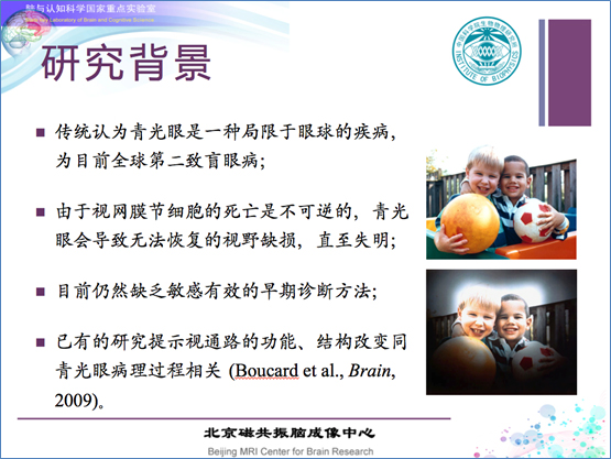
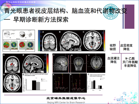
2. 和同仁医院合作关于“青光眼患者视皮层功能损害”的研究,率先将功能成像技术引入对青光眼(开角型)皮层功能损害神经机制的探索工作,成果发表于2010年的 Investigative Ophthalmology and Visual Science(IOVS,眼科学界基础研究杂志中排名 Top 2)。目前正在开展皮层功能损害的脑血流、代谢、结构等多重生物学指标的探索研究。

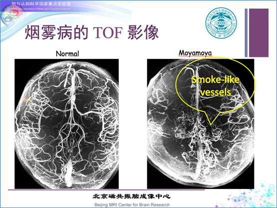
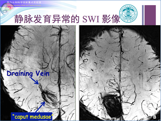
3. 和天坛医院、宣武医院合作开展超高场7T磁共振系统影像的临床应用研究,包括对超高信噪比和分辨率的大脑结构、颅内血管影像的认识和理解,基础知识库(磁共振联合解剖的图谱)的建立,以及患者影像资料的采集和临床诊断分析。
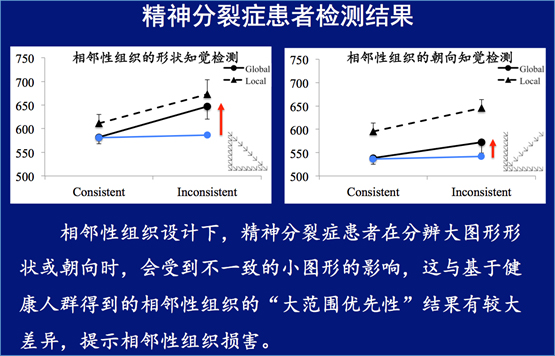
4. 和华西医院精神科,北京回龙观医院等单位合作开展"精神分裂症患者的视知觉组织改变"研究,试图探索特异而又稳定的精神疾病行为学指标,建立针对精神疾患、精神心理异常的敏感有效的认知科学检测方法。
1. 基金委重点项目,31730039,"基本情绪和视觉基本单元的相互作用及'大范围首先'的皮层下通路神经表达",2018/01-2022/12,315万元,在研,参加;
2. 科技部"973"项目子课题,2012CB825505,"认知基本单元研究的实验范式和心理病理学的结合",2012/01-2016/12,288万元,结题,参加;
3. 中国科学院战略性先导科技专项(B类),XDB02010001,"认知基本单元的大范围首先的神经表达:皮层下通路的脑功能联结图谱",2012/10-2016/12,880万元,结题,参加;
4. 基金委重大研究计划,91132302,"情感与视觉记忆:它们的相互作用及神经环路研究",2012/01-2015/12,300万元,结题,参加;
5. 科技部"973"项目子课题,2011CB504601,"与近视发生相关的视觉信息加工与处理异常 之环境因素影响及干预基础研究",2011/1-2015/12,288万元,结题,参加;
6. 科技部创新方法工作项目,2010IM030800,"神经外科患者术前脑功能区定位检测方法研究",2010/09-2013/8,180万元,结题,主持;
7. 基金委重大研究计划项目,90820307,"注意的认知机制和神经表达-创立和发展'大范围首先'的基于物体的注意理论",2009/1-2012/12,288万元,结题,参加;
8. 基金委重点项目,30830101,"功能磁共振成像和神经导航的微创神经外科学研究",200 9/1-2012/12,288万元,结题,参加;
9. 中国科学院知识创新方向性项目,KSCX2-YW-R-122,"大范围不变性知觉的神经表达:' 大范围首先'在颞叶的神经表达",2007/03-2010/02,288万元,结题,参加;
10. 科技部"973"项目子课题,2005CB522801,"特征捆绑和不变性知觉的脑认知功能成像",2005/1-2009/12,682.5万元,结题,参加;
11. 科技部"973"项目子课题,2004CB318101,"视觉注意的基本表达及其脑功能成像研究",2004/1-2008/12,288万元,结题,参加。
1. Deng, X., Wang, B.*, Zong, F., Yin, H., Yu, S., Zhang, D., ... & Zhang, Y.* (2021). Right‐hemispheric language reorganization in patients with brain arteriovenous malformations: A functional magnetic resonance imaging study. Human Brain Mapping. 1-14. https://doi.org/10.1002/hbm.25666
2. Wu, R., Wang, B.*, Zhuo, Y., & Chen, L. (2021). Topological dominance in peripheral vision. Journal of Vision, 21(10):19, 1-14. https://doi.org/10.1167/jov.21.10.19
3. Kang M.T., Wang B#, Ran A.R., Gan J.H., Du J.L., Mayinuer, Y., Liang X.T., Li S.M. & Wang N.L. (2021). Brain Activation Induced by Myopic and Hyperopic Defocus From Spectacles. Front. Hum. Neurosci., 15. https://doi.org/10.3389/fnhum.2021.711713
4. Xi, H., Wu, R., Wang, B., & Chen, L. (2020). Topological difference between target and flankers alleviates crowding effect. Journal of Vision, 20(9), 9. https://doi.org/10.1167/jov.20.9.9
5. Deng, X., Wei, X., Zhang, Y., Wang, B., Zhang, D., Yu, S., Jiang, T., & Zhao, J. (2020). Impact of AVM location on language cortex right-hemisphere reorganization: A voxel-based lesion-symptom mapping study. Clin Neurol Neurosurg, 189, 105628. https://doi.org/10.1016/j.clineuro.2019.105628
6. Meng, Q., Wang, B., Cui, D., Liu, N., Huang, Y., Chen, L., & Ma, Y. (2019). Age-related changes in local and global visual perception. Journal of Vision, 19(1), 10. https://doi.org/10.1167/19.1.10
7. Kong, Q. L., Zhang, Z. H., Yang, Q., Fan, Z. Y., Wang, B., An, J., & Zhuo, Y. (2019). 7T TOF-MRA shows modulated orifices of lenticulostriate arteries associated with atherosclerotic plaques in patients with lacunar infarcts. European Journal of Radiology, 118, 271-276. https://doi.org/10.1016/j.ejrad.2019.07.032
8. Sun, K. B., Cui, J. F., Wang, B., Jiang, T., Chen, Z. W., Cong, F., Zhuo, Y., Liang, S. L., Xue, R., Yu, X. G., & Chen, L. (2018). Magnetic resonance imaging of tuberous sclerosis complex with or without epilepsy at 7 T. Neuroradiology, 60(8), 785-794. https://doi.org/10.1007/s00234-018-2040-2
9. Deng, X., Zhang, D., Zhang, Y., Wang, R., Wang, B., & Zhao, J. (2017). Moyamoya disease with occlusion of bilateral vertebral arteries and the basilar artery fed by the collateral vessels of vertebral arteries: A rare case report. J Clin Neurosci, 42, 116-118. https://doi.org/10.1016/j.jocn.2017.03.043
10. Deng, X., Xu, L., Zhang, Y., Wang, B., Wang, S., Zhao, Y., Cao, Y., Zhang, D., Wang, R., Ye, X., Wu, J., & Zhao, J. (2016). Difference of language cortex reorganization between cerebral arteriovenous malformations, cavernous malformations, and gliomas: a functional MRI study. Neurosurg Rev, 39(2), 241-249. https://doi.org/10.1007/s10143-015-0682-7
11. Deng, X., Zhang, Z., Zhang, Y., Zhang, D., Wang, R., Ye, X., Xu, L., Wang, B., Wang, K., & Zhao, J. (2016). Comparison of 7.0- and 3.0-T MRI and MRA in ischemic-type moyamoya disease: preliminary experience. J Neurosurg, 124(6), 1716-1725. https://doi.org/10.3171/2015.5.JNS15767
12. Zhang, S., Wang, B.#, Xie, Y., Zhu, S., Thomas, R., Qing, G., Zhang, C., & Wang, N. (2015). Retinotopic Changes in the Gray Matter Volume and Cerebral Blood Flow in the Primary Visual Cortex of Patients With Primary Open-Angle Glaucoma. Invest Ophthalmol Vis Sci, 56(10), 6171-6178. https://doi.org/10.1167/iovs.15-17286
13. Deng, X. F., Zhang, Y., Xu, L., Wang, B., Wang, S., Wu, J., Zhang, D., Wang, R., Wang, J., & Zhao, J. Z. (2015). Comparison of language cortex reorganization patterns between cerebral arteriovenous malformations and gliomas: a functional MRI study. Journal of Neurosurgery, 122(5), 996-1003. https://doi.org/10.3171/2014.12.Jns14629
14. Lei, Q., Bao, Y., Wang, B., & Gutyrchik, E. (2012). fMRI correlates of inhibition of return in perifoveal and peripheral visual field. Cogn Process, 13 Suppl 1, S223-227. https://doi.org/10.1007/s10339-012-0487-3
15. Yu, S., Yan, L., Yao, Y., Wang, S., Yang, M., Wang, B., Zhuo, Y., Ai, L., Miao, X., Zhao, J., & Wang, D. J. (2012). Noncontrast dynamic MRA in intracranial arteriovenous malformation (AVM), comparison with time of flight (TOF) and digital subtraction angiography (DSA). Magn Reson Imaging, 30(6), 869-877. https://doi.org/10.1016/j.mri.2012.02.027
16. Yan, L., Zhuo, Y., Wang, B., & Wang, D. J. (2011). Loss of Coherence of Low Frequency Fluctuations of BOLD FMRI in Visual Cortex of Healthy Aged Subjects. Open Neuroimag J, 5, 105-111. https://doi.org/10.2174/1874440001105010105
17. Qing, G., Zhang, S., Wang, B., & Wang, N. (2010). Functional MRI signal changes in primary visual cortex corresponding to the central normal visual field of patients with primary open-angle glaucoma. Invest Ophthalmol Vis Sci, 51(9), 4627-4634. https://doi.org/10.1167/iovs.09-4834
18. Wang, B.*, Zhou, T. G., Zhuo, Y., & Chen, L*. (2007). Global topological dominance in the left hemisphere. Proc Natl Acad Sci U S A, 104(52), 21014-21019. https://doi.org/10.1073/pnas.0709664104
19. Liu, Z. X., & Wang, B. (2002). Full-screen ultrafast video modes over-clocked by simple VESA routines and registers reprogramming under MS-DOS. Behavior Research Methods Instruments & Computers, 34(2), 257-259. https://doi.org/Doi 10.3758/Bf03195453
(资料来源:王波副研究员,2021-11-02)
王波 博士 副研究员
研究方向:视知觉和(功能)磁共振成像技术的转化研究
电子邮件:bwang@ibp.ac.cn
电 话:010-64888565转809
通讯地址:北京市朝阳区大屯路15号(100101)
英文版个人网页:http://english.ibp.cas.cn/sourcedb/rck/EN_zkyqchhy/202005/t20200519_339617.html