Fluorescent image of BPAE cells
发布时间:2021-11-10
|
【 大 中 小 】
|
【打印】 【关闭】

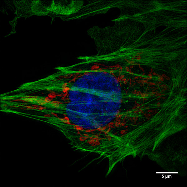
Fig1.BPAE cells
Courtesy of Bei Liu, Tao Xu'Group, CBI, IBP, CAS.
Image Details: Imaged by Delta Vision OMX Super-resolution Microscope
Mode: Structured Illumination Microscopy (SIM)
Scale bars: 5 μm
BPAE Cells With Mito Tracker Red CMXRos, Alexa Fluor 488 phalloidin, DAPI.

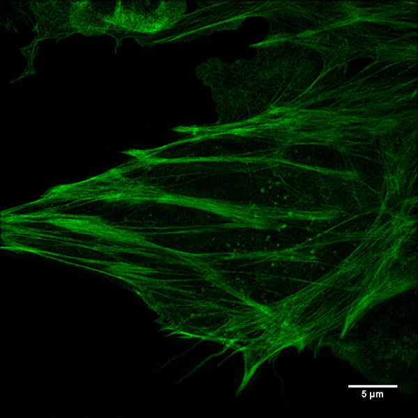
Fig1-1.BPAE cells
Courtesy of Bei Liu, Tao Xu'Group, CBI, IBP, CAS.
Image Details: Imaged by Delta Vision OMX Super-resolution Microscope
Mode: Structured Illumination Microscopy (SIM)
Scale bars: 5 μm
BPAE Cells With Alexa Fluor 488 phalloidin.

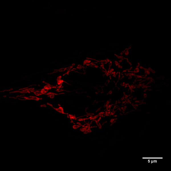
Fig1-2.BPAE cells
Courtesy of Bei Liu, Tao Xu'Group, CBI, IBP, CAS.
Image Details: Imaged by Delta Vision OMX Super-resolution Microscope
Mode: Structured Illumination Microscopy (SIM)
Scale bars: 5 μm
BPAE Cells With Mito Tracker Red CMXRos.

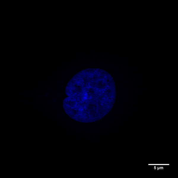
Fig1-3.BPAE cells
Courtesy of Bei Liu, Tao Xu'Group, CBI, IBP, CAS.
Image Details: Imaged by Delta Vision OMX Super-resolution Microscope
Mode: Structured Illumination Microscopy (SIM)
Scale bars: 5 μm
BPAE Cells With DAPI.


