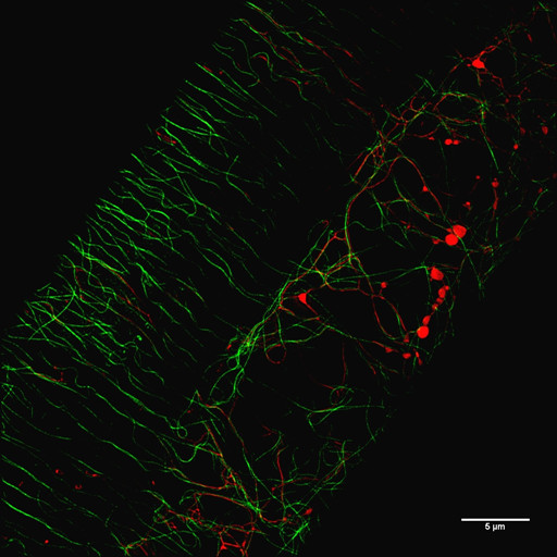
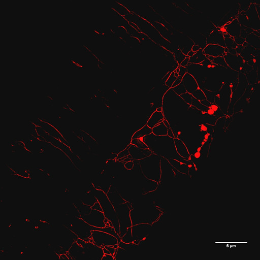
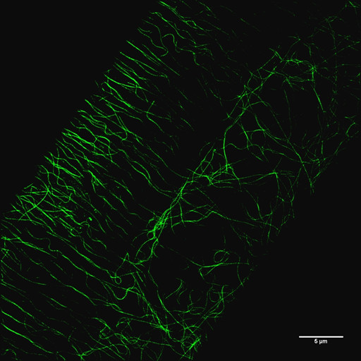
线虫表皮细胞内微管及溶酶体的结构光照明超分辨显微成像

fig1.Microtubule-GFP and Lysosomes-mCherry in the hypodermal cell of C.elegans

fig2.Lysosomes-mCherry in the hypodermal cell of C.elegans

fig3.Microtubule-GFP in the hypodermal cell of C.elegans
Courtesy of Yanwei Wu, Xiaochen Wang’s Group, NIBS
Image Details: Imaged by Delta Vision OMX Super-resolution Microscope
Mode: Structured Illumination Microscopy (SIM)
Scale bars: 5 μm