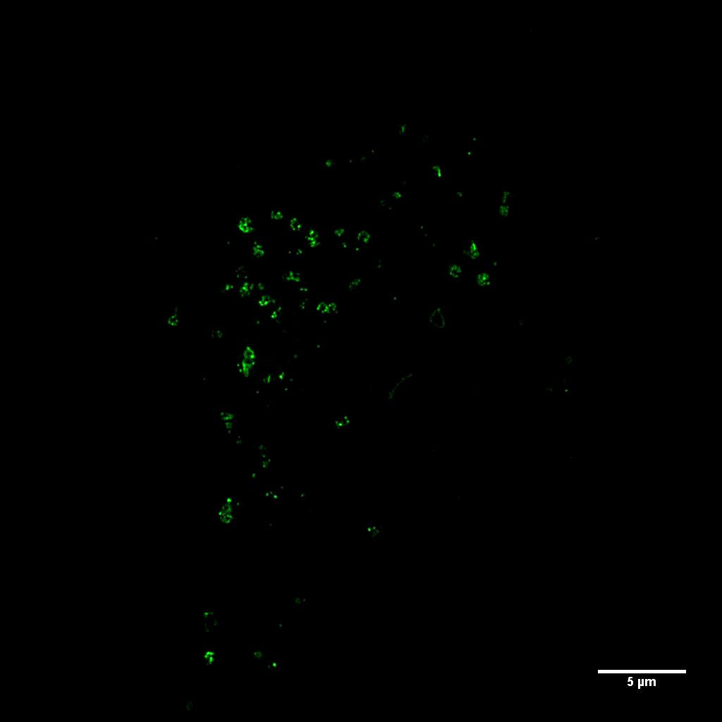
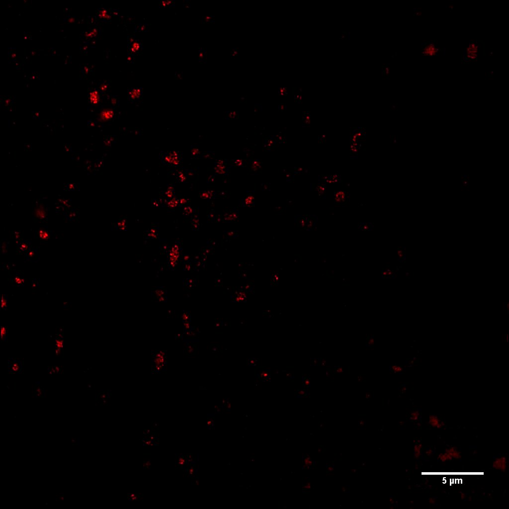
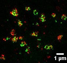
海拉细胞免疫标记LC3的结构照明超分辨显微成像


Fig1. Hela cells transfected with WIPI1-GFP and immunostained with antibody against LC3 (red) were analysed by 3DSIM
Courtesy of Yan Zhao, Hong Zhang’s Group, IBP, CAS.
Image Details: Imaged by Delta Vision OMX Super-resolution Microscope
Mode: Structured Illumination Microscopy (SIM)
Scale bars: 5 μm